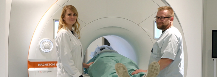
Magnetic Resonance Imaging (MRT)
The abbreviation MRI stands for magnetic resonance imaging. MRI uses strong, alternating magnetic fields and radio waves of varying strength to image the inside of the body.
MRI is a cross-sectional imaging technique without the use of X-rays and is used primarily for detailed imaging of the brain, spinal cord, spine, vessels and joints.
Examination procedure::
You lie on an examination table in the MRI unit during the examination. The modern MRI machines are open on both sides and have their own ventilation. Since it is technically very loud during the examination, you will be provided with earplugs and headphones as hearing protection. You can communicate with us at any time. On average, an MRI examination lasts 30 minutes, in rare cases or in complicated situations - a little longer. Whether a contrast medium has to be administered via the vein is decided individually depending on the problem. Before the examination, you will be informed about the exact procedure and any necessary administration of contrast media.
What do you need to pay attention to?
Since the MRI machine generates a strong magnetic field, all metallic objects must be removed before the examination. This includes coins, check cards, cell phones, piercings and jewelry, removable dentures, hearing aids, belts. Implanted metallic objects, such as pacemakers, event recorders, artificial heart valves, pain pumps, intracranial stimulation electrodes, etc., are partially not or only partially MRI-capable.
It must therefore be checked in advance whether and under what conditions an MRI examination is possible.
Please inform your referring physician in advance about these implants and be sure to bring your implant ID card to the examination.


